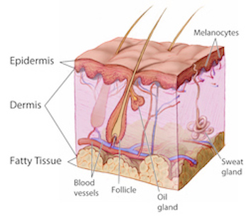
 There are three main types of skin biopsies: shave, punch and excision. According to PRP patients and caregivers, shave biopsies and punch biopsies are the types used as a PRP diagnostic tool.
SHAVE BIOPSY
❏½ A doctor uses a tool similar to a razor to remove a small section of the top layers of skin (epidermis and a portion of the dermis).
❏½ No stitches are required. The wound forms a scab that should heal in 1–3 weeks.
❏½ As a shave biopsy does not include the full thickness of the skin, the drawback of such a biopsy is that it may be difficult for a pathologist to rule out or identify invasive disease.
❏½ After a local anesthetic is injected, a surgical knife (scalpel) is used to shave off the growth . Stitches are not needed.
❏½ Any bleeding can usually be controlled with a chemical that stops bleeding and by applying pressure.
❏½ The biopsy site is then covered with a bandage or sterile dressing.
PUNCH BIOIPSY
❏½ The punch biopsy is generally the most useful type of biopsy. It is quick to perform, convenient, and only produces a small wound. It creates a full thickness sample of skin that allows the pathologist to get a good overview of the epidermis, dermis, and most of the time, the subcutis also.
❏½ In a punch biopsy, a cookie cutter-like, circular tool to remove a small section of skin including deeper layers
❏½ A disposable skin biopsy punch is used, which has a round stainless steel blade ranging from 2–6 mm in diameter. The 3 and 4 mm punches are the most common sizes used.
❏½ The clinician holds the instrument perpendicular to the anaesthetized skin and rotates it to pierce the skin. Using a forceps and scissors the skin sample is subsequently removed.
❏½ After a local anesthetic is injected, a small, sharp tool that looks like a cookie cutter (punch) is placed over the lesion, pushed down, and slowly rotated to remove a circular piece of skin .
❏½ The skin sample is lifted up with a tool called a forceps or a needle and is cut from the tissue below.
❏½ A suture may be used to close a punch biopsy wound or help control bleeding if necessary. However, if the wound is small, it may heal adequately without it. Stitches may not be needed for a small skin sample. If a large skin sample is taken, one or two stitches may be needed. Pressure is applied to the site until the bleeding stops.
❏½ The wound is then covered with a bandage or sterile dressing.
EXCISION BIOPSY
❏½ Excision biopsy refers to complete removal of a skin lesion, such as a skin cancer in which a margin of surrounding skin is taken to improve chances of complete removal. Smaller lesions are most often removed using a scalpel blade as an ellipse, with primary closure using sutures. Larger excisions may be repaired using a skin flap (moving adjacent skin to cover the wound) or graft (skin taken from another site to patch the wound).
❏½ This type of biopsy may be useful to provide a better overview for the pathologist, which can improve diagnostic accuracy. It can also be useful when deeper layers or tissue are believed to be involved in the disease process (eg, subcutaneous fat or medium-sized blood vessels).
❏½ A doctor uses a small knife (scalpel) to remove an entire lump or an area of abnormal skin, including a portion of normal skin down to or through the fatty layer of skin
❏½ After a local anesthetic is injected, the entire lesion is removed with a scalpel. Stitches are used to close the wound. Pressure is applied to the site until the bleeding stops. The wound is then covered with a bandage or sterile dressing. If the excision is large, a skin graft may be needed.
SOURCES
❏½ http://www.dermnetnz.org/topics/skin-biopsy/
❏½ http://www.webmd.com/cancer/skin-biopsy#1
❏½ http://www.mayoclinic.org/tests-procedures/skin-biopsy/home/ovc-20196287
❏½ http://www.webmd.com/cancer/what-is-a-biopsy#1
❏½ http://www.medicinenet.com/skin_biopsy/article.htm

